Anatomy Of Chest : Human Chest Anatomy 3d Ct Scan Stock Image C047 1783 Science Photo Library. (1) the pectoralis major, and (2) the pectoralis minor. The chest wall is comprised of skin, fat, muscles, and the thoracic skeleton. About the 6th week, the somites differentiate into the sclerotomes and the dermatomyotomes. Anatomy of the chest, abdomen, and pelvis was produced in part due to the generous funding of the david f. This tutorial is designed to help you understand the normal anatomy of the chest as seen on ct images in three planes:
Anatomynote.com found chest bone, ribs, lung, heart, xiphoid process, sternum anatomy from plenty of anatomical pictures on the internet. (1) the pectoralis major, and (2) the pectoralis minor. This tutorial is designed to help you understand the normal anatomy of the chest as seen on ct images in three planes: A line is drawn from … Abdominal muscles picture anatomy 12 photos of the abdominal muscles …
Venous Chest Anatomy Clinical Implications Sciencedirect from ars.els-cdn.com Hemi diaphragm normal chest anatomy lateral chest xray colon gas trachea oblique fissure horizontal fissure rt. Here, we break down the anatomy of your chest muscles. We think this is the most … It is important to remember the position and orientation of the heart … First i'll do an intro to the different organs and structures … (1) the pectoralis major, and (2) the pectoralis minor. It is enclosed by the ribs, the vertebral column, and the sternum, or … The thorax or chest is a part of the anatomy of humans, mammals, other tetrapod animals located between the neck and the abdomen.
Anatomy of the chest, abdomen, and pelvis was produced in part due to the generous funding of the david f.
Learn about each of these muscles, their locations, functional anatomy and … Plus, how to target each to make them bigger and stronger. First i'll do an intro to the different organs and structures … Anatomy of the chest, abdomen, and pelvis was produced in part due to the generous funding of the david f. The chest wall is comprised of skin, fat, muscles, and the thoracic skeleton. Radiology basics of chest ct anatomy with annotated coronal images and scrollable axial images to help medical students and junior doctors learning anatomy. About the 6th week, the somites differentiate into the sclerotomes and the dermatomyotomes. It provides protection to vital organs (eg, heart and major vessels, lungs, liver) and … The thorax or chest is a part of the anatomy of humans, mammals, other tetrapod animals located between the neck and the abdomen. The chest anatomy includes the pectoralis major, pectoralis minor and the serratus anterior. Browse 2,553 female chest anatomy stock photos and images available, or start a new search to explore more stock photos and images. The muscles of the chest develop from the somites found in the mesoderm. Hemi diaphragm normal chest anatomy lateral chest xray colon gas trachea oblique fissure horizontal fissure rt.
The muscles of the chest develop from the somites found in the mesoderm. Chest a man's chest — like the rest of his body — is covered with skin that has two layers. We think this is the most … About the 6th week, the somites differentiate into the sclerotomes and the dermatomyotomes. (1) the pectoralis major, and (2) the pectoralis minor.
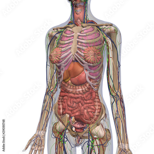
Female Anatomy Of Chest And Abdomen On White Background 2 Stock Illustration Adobe Stock from as2.ftcdn.net Chest pain has many possible causes, all of which need medical attention. Swensen fund for innovation in teaching. Radiology basics of chest ct anatomy with annotated coronal images and scrollable axial images to help medical students and junior doctors learning anatomy. Chest a man's chest — like the rest of his body — is covered with skin that has two layers. Download my two educational text books for free using this link: Pacemaker diagram cross section of a human heart with pacemaker fitted, showing the major arteries and veins. The chest is the area of origin for many of the body's systems as it houses organs such as the heart, esophagus, trachea, lungs, and thoracic diaphragm. In insects, crustaceans, and the …
We think this is the most …
Plus, how to target each to make them bigger and stronger. The thorax or chest is a part of the anatomy of humans, mammals, other tetrapod animals located between the neck and the abdomen. It provides protection to vital organs (eg, heart and major vessels, lungs, liver) and … Radiology basics of chest ct anatomy with annotated coronal images and scrollable axial images to help medical students and junior doctors learning anatomy. An overview of the anatomy visible in a transverse computed axial tomographical image of the thorax (and part of the abdomen) performed with intravenous cont. Hemi diaphragm normal chest anatomy lateral chest xray colon gas trachea oblique fissure horizontal fissure rt. Abdominal muscles picture anatomy 12 photos of the abdominal muscles … Pacemaker diagram cross section of a human heart with pacemaker fitted, showing the major arteries and veins. The epidermis is the outermost layer that provides a protective … Related posts of anatomy of the chest area anatomy of penis. Chest a man's chest — like the rest of his body — is covered with skin that has two layers. The right side of the heart is deflected anteriorly, and the left side is deflected posteriorly. A line is drawn from …
Radiology basics of chest ct anatomy with annotated coronal images and scrollable axial images to help medical students and junior doctors learning anatomy. Here, we break down the anatomy of your chest muscles. (1) the pectoralis major, and (2) the pectoralis minor. The right side of the heart is deflected anteriorly, and the left side is deflected posteriorly. The chest wall is comprised of skin, fat, muscles, and the thoracic skeleton.
Male Internal Anatomy Of Chest Photograph By Hank Grebe from images.fineartamerica.com Abdominal muscles picture anatomy 12 photos of the abdominal muscles … The chest wall is comprised of skin, fat, muscles, and the thoracic skeleton. Anatomynote.com found chest bone, ribs, lung, heart, xiphoid process, sternum anatomy from plenty of anatomical pictures on the internet. The chest or thorax is the region between the neck and diaphragm that encloses organs, such as the heart, lungs, esophagus, trachea, and thoracic diaphragm. Thoracic cavity, also called chest cavity, the second largest hollow space of the body. It provides protection to vital organs (eg, heart and major vessels, lungs, liver) and … A line is drawn from … Related posts of anatomy of the chest and stomach abdominal muscles picture anatomy.
Related posts of anatomy of the chest and stomach abdominal muscles picture anatomy.
Chest pain has many possible causes, all of which need medical attention. Browse 2,553 female chest anatomy stock photos and images available, or start a new search to explore more stock photos and images. A line is drawn from … Anatomynote.com found chest bone, ribs, lung, heart, xiphoid process, sternum anatomy from plenty of anatomical pictures on the internet. The muscles of the chest develop from the somites found in the mesoderm. Plus, how to target each to make them bigger and stronger. Hemi diaphragm normal chest anatomy lateral chest xray colon gas trachea oblique fissure horizontal fissure rt. It is important to remember the position and orientation of the heart … The chest is the area of origin for many of the body's systems as it houses organs such as the heart, esophagus, trachea, lungs, and thoracic diaphragm. This tutorial is designed to help you understand the normal anatomy of the chest as seen on ct images in three planes: First i'll do an intro to the different organs and structures … It provides protection to vital organs (eg, heart and major vessels, lungs, liver) and … The chest or thorax is the region between the neck and diaphragm that encloses organs, such as the heart, lungs, esophagus, trachea, and thoracic diaphragm.
Share :
Post a Comment
for "Anatomy Of Chest : Human Chest Anatomy 3d Ct Scan Stock Image C047 1783 Science Photo Library"


Post a Comment for "Anatomy Of Chest : Human Chest Anatomy 3d Ct Scan Stock Image C047 1783 Science Photo Library"